


Core needle biopsy (or core biopsy) is performed by inserting a small hollow needle through the skin and into the organ or abnormality to be investigated. The needle is then advanced within the cell layers to remove a sample or core. Needle biopsy is also a type of percutaneous (through the skin) biopsy. The needle may be designed with a cutting tip to help remove the sample of tissue. Core biopsy is often performed with the use of spring loaded gun to help remove the tissue sample.
Core biopsy is typically performed under image guidance such as CT imaging, ultrasound or mammography. The needle is either placed by hand or with the assistance of a sampling device. Multiple insertions are often made to obtain sufficient tissue, usually multiple samples are taken. Patients may experience a slight pressure, but usually do not experience significant pain. As tissue samples are taken, a click may be heard from the sampling instrument. The core tissue samples will be sent to the pathology laboratory for diagnosis. In some cases, the pathologist can attend the biopsy to examine imprints of the samples with a microscope. This can allows the pathologist to determine the adequacy of the sample and perhaps offer preliminary results.
Learn more about core needle biopsy on the breast.
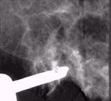
This mammogram image shows the vacuum assisted breast biopsy probe (Biopsys Mammotome) is positioned in the breast and aligned with calcifications (white specs above needle opening)
Core biopsy is sometimes suction assisted with a vacuum device. This method enables to removal of multiple samples with only one needle insertion. Vacuum assisted core biopsy is being used more and more in breast biopsy procedures and is guided via stereotactic mammography or ultrasound imaging. However, unlike core biopsy, the vacuum assisted biopsy probe is inserted just once into the tissue through a tiny skin nick. Multiple samples are then taken using a rotation of the sampling needle aperture (opening) and with the assistance of suction. Click here for more information on vacuum assisted breast biopsy.
Endoscopic biopsy is a very common type of biopsy that is done through an endoscope (a fiber optic cable for viewing inside the body) which is inserted into the body along with sampling instruments. The endoscope allows the physician to visualize the abnormality and guide the sampling. Endoscopic biopsy may be performed of the gastrointestinal tract (alimentary tract endoscopy), urinary bladder (cystoscopy), abdominal cavity (laparoscopy), joint cavity (arthroscopy), mid-portion of the chest (mediastinoscopy), or trachea and bronchial system (laryngoscopy and bronchoscopy), either through a natural body orifice or a small surgical incision. The endoscopist can directly visualize an abnormal area on the lining of the organ in question and pinch off tiny bits of tissue with forceps attached to a long cable that runs inside the endoscope.
Punch biopsy is typically used by dermatologists to sample skin rashes, moles and other small masses. After a local anesthetic is injected, a biopsy punch, which is similar in function to a small (3 mm to 4 mm or 0.15 inch in diameter) version of a cookie cutter, is used to cut out a cylindrical piece of skin. The opening is typically closed with a suture (small stitches) and heals with minimal scarring. Punch biopsy may also be performed when removing small tissue samples from the cervix.
Surface biopsy involves sampling or scraping the surface of a sore or tumor to remove cells for pathologic testing. Surface biopsy is often performed by dermatologists to remove a small piece of skin to test for carcinoma (cancerous tissue).
Surgical biopsy can be excisional (removal of an entire lesion) or incisional (removal of a piece of a lesion). Until about a decade ago, most biopsies performed were open surgical procedures. However, surgical biopsy is less common now. Surgical biopsies can be performed on abnormalities that can be seen or felt by the surgeon or pre-operative imaging can help provide a road map to the lesion. In cases of non-palpable breast lesions, a percutaneous wire can be placed in or near the lesion using mammogram or ultrasound for guidance. This marker wire provides a target for the surgeon. The removed tissue is then histologically analyzed by a pathologist (a special laboratory physician uses microscopic analysis of the tissue to determine its type).
Learn more about open surgical biopsy on the breast.
Some types of tumors (such as lymphoma, a cancer of the lymphocyte blood cells) need to be examined whole to allow an accurate diagnosis. Thus enlarged lymph nodes are good candidates for excisional biopsy.
Surgical biopsy has some disadvantages versus percutaneous needle biopsy. Surgical biopsies require sutures (stitches) and can leave a disfiguring scar, they carry a small risk of mortality (due to risks of anesthesia) and moderate chances of bleeding, infection, wound healing problems and even fracture or migration of the localizing wire. Surgical biopsy usually requires one day of recuperation at home and usually is significantly more expensive than the alternative methods. Advantages of surgical biopsy include the ability to usually obtain a larger sample or complete removal of a lesion versus percutaneous methods. Bleeding encountered during a surgical procedure is easier to control than with percutaneous methods.
