


- Related Articles
- What is Mammography?
- How is Mammography Performed?
- Views Taken During Screening and Diagnostic Mammography
- Breast Compression During Mammography
- Minimizing Pain and Discomfort During Mammography
- Skin Markers During Mammography
- Special Mammography Techniques:
- Additional Information on Mammography
For screening mammography each breast is imaged separately:
- typically from above (cranial-caudal view, CC) and
- from an oblique or angled view (mediolateral-oblique, MLO)
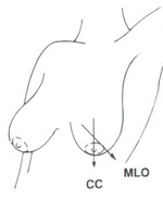
Cranio-caudal (CC) view and mediolateral oblique (MLO) mammographic view
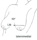
Latero medial (LM) mammographic view
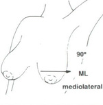
Medio lateral (ML) mammographic view
For diagnostic mammography, each breast is imaged separately:
- from above (cranial-caudal view, CC)
- from an oblique or angled view (mediolateral-oblique, MLO) and
- supplemental views tailored to the specific problem are often performed. These can include views from each side (lateromedial, LM: from the outside towards the center and mediolateral view, ML: from the center of the chest out), exaggerated cranial-caudal, magnification views, spot compression, and others.
- if screening mammography has been performed first and the resulting CC and MLO views are of sufficient quality, they may not need to be repeated if diagnostic mammography is required.
A cleavage view (also called "valley view") is a mammogram view that images the most medial (central) portions of the breasts. This is the portion of breast tissue "in the valley" between the two breasts. When one breast is imaged and the other breast is left out of the compression field, some of the breast being imaged may get pulled or left out too. To get as much medial tissue as possible, the mammogram technologist will place both breasts on the plate at the same time to image the medial half of both breasts.
A cleavage view may be performed when there is a questionable density on the medial edge of the mammogram film and the radiologist needs to see more of this density (if possible). A cleavage view may also be performed if the radiologist sees something suspicious in the mediolateral-oblique (MLO) mammogram view and cannot find the area on the cranial-caudal view (CC) view.
Detailed information on mammography positioning and imaging.
Breast compression is necessary to flatten the breast so that the maximum amount of tissue can be imaged and examined. Breast compression may cause some discomfort, but it only lasts for a brief time during the mammography procedure. Patients should feel firm pressure due to compression but no significant pain. If you feel pain, please inform the technologist. During the mammography examination, breast compression should only be applied two to four times per breast for a few seconds each time (see below for description of views taken during screening and diagnostic mammography).
Breast compression is necessary during mammography in order to:
- Flatten the breast so there is less tissue overlap for better visualization of anatomy and potential abnormalities. For example, inadequate compression can lead to poor imaging of microcalcifications, tiny calcium deposits that are often an early sign of breast cancer.
- Reduce overlapping normal shadows, which can appear as suspicious regions on the film.
- Allow the use of a lower x-ray dose since a thinner amount of breast tissue is being imaged
- Immobilize the breast in order to eliminate image blurring caused by motion
- Reduce x-ray scatter which also leads to image degradation
Some mammography facilities will allow the patient to control the breast compression herself during mammography. See the section below on Minimizing Pain and Discomfort During Mammography for more information.
